BERKOWITZ LAB
School of Medicine
Department of Ophthalmology, Visual and Anatomical Sciences
Detroit, MI
baberko@med.wayne.edu
(313) 577-9305

BERKOWITZ LAB


|
Anterior cardinal veins
|
These bilaterally symmetrical, paired veins drain blood from the head and neck into their respective common cardinal veins during the early 4th week. Regression of the proximal end of the left anterior cardinal vein and formation of
the left brachiocephalic vein at the end of the second month results in all blood from the head and neck flowing into the proximal end of the right anterior cardinal which thus forms the superior vena cava. The distal ends of the anterior
cardinals give rise to the internal jugular veins. The designation of these veins as anterior is a misnomer. They should be referred to as superior cardinal veins in the bipedal human.
|
|
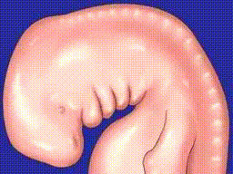
Aortic arches
|
These paired arteries are derived from a basketwork of arteries surrounding the pharynx of piscine progenitors. In fish, they vascularize the gill bars and function to extract oxygen from water flowing through the pharynx. The
aortic arches in humans correspond to arches 1, 2, 3, 4, and 6 of the ancestral fish but have become highly modified to form the great vessels of the thorax.
|
|
Aortic sac
|
The aortic arches (2, 3, 4, and 6) sprout from this most distal specialization of the truncus arteriosus. Its derivatives include the proximal end of the arch of the aorta and the brachiocephalic artery.
|
|
Aortic valve
|
See semilunar valves.
|
|
Ascending aorta
|
This vessel is the most proximal segment of the aorta, derived from the truncus arteriosus.
|
|
Atrioventricular node (AVN)
|
This secondary pacemaker region within the superior endocardial cushion receives impulses from the sinoatrial node (SAN) and controls the beating of the two ventricles.
|
|
Atrioventricular sulcus
|
This constriction first demarcates the boundary between the primitive atrium and primitive ventricle at the end of the 3rd week.
|
|
Atrioventricular valves
|
These valves prevent backflow into the atria during systole. They are sculpted from ventricular muscle. Their cusps or leaflets are tethered to papillary muscles of the ventricular wall by tough sinews called chordae tendinae. The atrioventricular
valve of the left ventricle, the bicuspid or mitral valve, is characterized by two sets of cusps, chordae tendinae, and papillary muscles while the tricuspid valve of the right ventricle normally possesses three.
|
|
Bicuspid (mitral) valve
|
See atrioventricular valves.
|
|
Bulboventricular sulcus
|
This constriction first demarcates the superior end of the primitive ventricle (presumptive left ventricle) from the inferior end of the bulbus cordis (presumptive right ventricle) at the end of the third week.
|
|
Bulbus cordis
|
This distinct segment of the primitive heart tube is first apparent at the end of the third week. Its inferior end will form much of the right ventricle while its superior end will first form an outflow channel called the conotruncus,
which will give rise to the more proximal conus cordis and the more distal truncus arteriosus.
|
|
Bundle of His
|
This collection of specialized myocytes forms a conduction pathway that carries electrical impulses from the atrioventricular node (AVN) to the muscular ventricular septum. It bifurcates on the septal ridge, sending one branch to the
right ventricle and another to the left ventricle.
|
|
Cardiac jelly
|
This acellular secretion of the myocardium plays a central role in septation of the heart and formation of the atrioventricular canals. During the early stages of heart development, the cardiac jelly comprises most of the thickness of
the heart wall. As the heart differentiates, however, the cardiac jelly disappears and myocardium soon accounts for the major proportion of the heart wall.
|
|
Cardiogenic region
|
This horshoe-shaped region lateral and cranial to the neural plates that will form the brain, will give rise to the lateral endocardial tubes and hence the primitive heart tube, by vasculogenesis.
|
|
Chordae tendinae
|
See "atrioventricular valves" above.
|
|
Common cardinal veins
|
The right and left common cardinal veins are short vessel segments which dump blood into the right and left horns of the sinus venosus respectively. They represent the confluence of the anterior and posterior cardinal veins.
|
|
Conduction system
|
Myocytes within the developing heart tube develop a more rapid rate of spontaneous depolarization to produce two pacemaker regions, the sinoatrial node (regulating atrial rhythm) and an atrioventricular node connected by a myocardial
conduction pathway (most notably within the crista terminalis) to coordinate regulate ventricular rhythmicity through the bundle of His. The primitive conduction system may develop as early as day 22.
|
|
Conotruncus
|
This distal region of the bulbus cordis will give rise to outflow regions of the ventricles themselves (the conus cordis) and the truncus arteriosus which will be divided into proximal ends of the ascending aorta and pulmonary trunk.
|
|
Conus cordis
|
This segment of the outflow tract is derived from the conotruncus and is remodeled to form outflow regions of the definitive left and right ventricles.
|
|
Coronary sinus
|
This vestige of the left horn of the sinus venosus becomes completely detached from the left anterior cardinal vein. Vessels deep to the epicardium will join with sprouts that grow from the coronary sinus to form the venous system that
will drain the myocardium itself.
|
|
Crista terminalis
|
This ridge of tissue demarcates the boundary between the sinus venarum (that part of the right atrium formed by intussusception of the right sinus venosus and the right lateral region of the primitive atrium, which forms the vestigial
right auricle. It contains a major conduction pathway connecting the sinoatrial node with the atrioventricular node.
|
|
Definitive left atrium
|
This definitive heart chamber is formed by intussusception of the pulmonary veins into the posterior wall of the primitive atrium.
|
|
Definitive left ventricle
|
Most of the definitive left ventricle is derived from the primitive ventricle, but part of it arises from the left wall of the conus cordis.
|
|
Definitive right atrium
|
This definitive heart chamber is formed by intussusception of the sinus venosus into the posterior wall of the primitive atrium.
|
|
Definitive right ventricle
|
This definitive chamber of the heart is mainly derived from the inferior end of the bulbus cordis, although parts of it may be derived from the right wall of the conus cordis and part of the primitive ventricle.
|
|
Dextrocardia
(cardiac heterotaxia) |
This term describes the condition resulting from an opposite, "dextral" folding (the apex of the "left" ventricle is now displaced to the right) of the heart, in contrast to the normal sinistral folding that occurs at the beginning of
the 4th week. This heart is a mirror-image of the orthotopic heart and may occur in cases of situs inversus.
|
|
Dorsal mesocardium
|
This dorsal mesentery of the primitive heart tube is ruptured in the 4th week to give rise to the transverse pericardial sinus of the adult heart.
|
|
Endocardial cushions
|
These thickenings in the atrioventricular canal are regions of thickened cardiac jelly.
|
|
Endocardium
|
The endocardium or lining of the heart tube is derived from the lateral endocardial tubes. It consists of endothelial and a thin subendothelial connective tissue.
|
|
Epicardium
(visceral pericardium) |
This outer tunic of thin serous membrane covers the myocardium and the coronary vessels. It is derived from a population of splanchnopleuric mesoderm distinct from the which forms the myocardium.
|
|
Foramen ovale
|
This inferior opening in the septum secundum allows blood to flow from the right to the left atrium during embryonic and fetal life.
|
|
Fossa ovalis
|
After birth, as pressure rises in the left ventricle, the septum primum is forced against the thicker septum secundum. In most cases the two septa physically fuse leaving evidence of the former foramen ovalis as a shallow depression,
the fossa ovalis, when viewed from within the right atrium.
|
|
Inferior vena cava
|
|
|
Lateral endocardial tubes
|
These paired tube develop within the lateral regions of the cardiogenic area by vasculogenesis. During cephalic and lateral flexion, they swing into the ventral thoracic region and fuse together to form the primitive heart tube. Cells
of the lateral endocardial tubes will give rise to the endocardium of the heart.
|
|
Left brachiocephalic vein
|
As the left anterior cardinal vein loses its connection with the left horn of the sinus venosus (presumptive coronary sinus), a new vein develops from thymic and thymus veins to connect the distal end of the left anterior cardinal vein
with the right anterior cardinal vein. The development of this left brachiocephalic vein allows the drainage of all blood from the left upper extremity and head and neck into the right atrium.
|
|
Membranous
ventricular septum |
The membranous ventricular septum develops from migrating neural crest ectomesenchymal cells that first form thickened truncoconal septa. These septa first form at the junction of the conus cordis and truncus arteriosus but zipper together
both superiorly and inferiorly to simultaneously separate the left and right ventricles and their respective outflow tracts. Their spiral growth, followed by cleavage of the aortic and pulmonary outflow channels along the plane of
the membranous septum, results in the characteristic twisting of the ascending aorta and pulmonary trunk about each other. Many heart malformations may arise completely, or in part, through defects in development of the membranous
ventricular septum, including ventricular septal defects, persistent truncus arteriosus, aortic or pulmonary stenosis, and tetralogy of Fallot.
|
|
Muscular
ventricular septum |
Septation of the ventricles is initiated by development and growth of a thickened ridge of musculature as the inferior part of the muscular wall of the bulboventricular sulcus protrudes into the lumen of the heart tube. The anterior
trabeculated part is called the primary ventricular fold (septum) and the smooth posterior region of the septum is called the inlet septum. The boundary between them is demarcated by a prominent trabeculation called the septomarginal
trabecula or moderator band. Growth of the muscular septum stops short, in the middle of the 7th week of completely separating the left and right ventricular chambers.
|
|
Myocardium
|
The myocardium, or cardiac muscle arises from a block of splanchnopleuric mesoderm that completely invests the primary heart tube. Myocardial cells secrete the cardiac jelly and some of them differentiate to form the conduction system
of the heart.
|
|
Oblique vein of the left atrium
|
This small vein draining the wall of the left atrium, arises from the distal end of the left sinus horn and is directly connected with the coronary sinus.
|
|
Ostium primum
|
This inferior opening in the septum primum closes completely as the septum primum fuses with the septum intermedium.
|
|
Ostium secundum
|
The ostium secundum develops within the superior region of the septum primum by the coalescence of cell death foci as the ostium primum closes.
|
|
Papillary muscles
|
These muscular protrusions of the ventricular walls anchor the chordae tendinae of the atrioventricular valves.
|
|
Posterior cardinal veins
|
More appropriately called inferior cardinal veins in humans, these bilateral, paired vessels drain the trunk and lower and extremities between the 4th and 8th weeks, when they are superseded by the subcardinal and supracardinal systems.
Distal elements and median anastomoses of the posterior cardinal system contribute small distal segments to the inferior vena cava and common iliac veins.
|
|
Primitive atrium
|
The primitive atrium appears as a small expansion of the primitive heart tube in the early 4th week. On the right side, it is superseded by intussusception of the right sinus venosus and is pushed laterally and anteriorly to form the
vestigial trabeculated right auricle. On the left side, it is superseded by intussusception of the pulmonary veins and is pushed laterally and anteriorly to form the vestigial trabeculated left auricle.
|
|
Primitive heart tube
|
The primitive heart tube is formed by fusion of the lateral endocardial tubes as they are brought into the ventral thoracic region by cephalic and lateral folding.
|
|
Primitive ventricle
|
This expansion of the primitive heart tube is apparent early in the 4th week. It will mainly form the definitive left ventricle, although a small part of it may contribute to development of the definitive right ventricle.
|
|
Pulmonary trunk
|
This outflow tract of the right ventricle develops from the truncus arteriosus.
|
|
Pulmonary valve
|
See semilunar valves.
|
|
Semilunar valves
|
These valves of the aortic and pulmonary outflow tracts (the aortic and pulmonary valves respectively) arise as cell death foci sculpt cusps from the left and right truncoconal septa (forming right and left cusps of both valves) and
from a minor anterior truncus swelling (a cusp of the pulmonary valve) and a minor posterior swelling (a cusp of the aortic valve).
|
|
Septum intermedium
|
This structure divides the single atrioventricular canal into right and left atrioventricular canals as the growing edges of the superior and inferior endocardial cushions meet and fuse during the 6th week. This septum provides a base
upon which the ventricular and atrial septa can fuse to completely separate the right and left ventricular chambers from each other and the right and left atrial chambers from each other respectively. Endocardial cushion defects with
concomitant atrial and ventricular septal defects may result from failure of complete fusion of the superior and inferior endocardial cushions and failure of the septum intermedium to form.
|
|
Septum primum
|
This membranous atrial septum forms adjacent to the left atrium. It first contains an ostium primum, which closes when the septum fuses with the septum intermedium, and then an ostium secundum which forms in it superior region as several
cell death foci coalesce.
|
|
Septum secundum
|
As the septum primum completes its development in the 6th week, a second, thicker septum grows from the atrial roof, just to the right of the septum primum. Growth of this septum secundum is incomplete resulting in the formation of a
formaen ovale in its inferior edge.
|
|
Sinus venarum
|
This is the smooth walled region of the definitive right atrium formed by intussusception of the right horn of the sinus venosus.
|
|
Sinus venosus
|
This chamber at the inferior end of the primitive heart tube is the site of confluence of left and right common cardinal, vitelline, and umbilical veins prior to remodeling of the inflow region of the heart. The left horn of the sinus
venosus is remodeled to form the coronary sinus and the right horn expands to form the sinus venarum, forming the definitive right atrium.
|
|
Superior vena cava
|
The superior vena cava arises from the proximal end of the right anterior cardinal vein.
|
|
Tricuspid valve
|
See atrioventricular valves.
|
|
Truncoconal septa
|
These septa of the outflow tracts are formed from neural crest ectomesenchymal cells which migrate into the truncus arteriosus through the 4th and 6th aortic arches. Their fusion simultaneously separates the right and left ventricular
chambers from one another along with their respective outflow tracts, the pulmonary trunk and aorta.
|
|
Truncus arteriosus
|
This distal segment of the bulbus cordis is divided into ascending aorta and pulmonary trunk by fusion of the truncoconal septa between the 5th and 9th weeks.
|